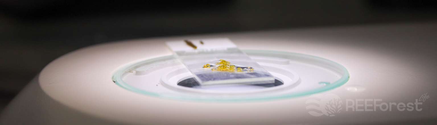
To ensure the success of marine restoration, it is important to start the process at the right time. That’s why we monitor the maturity of Cystoseira sensu lato‘s reproductive structures.
The time of year when the apices are fertile can vary greatly, both among locations and species.
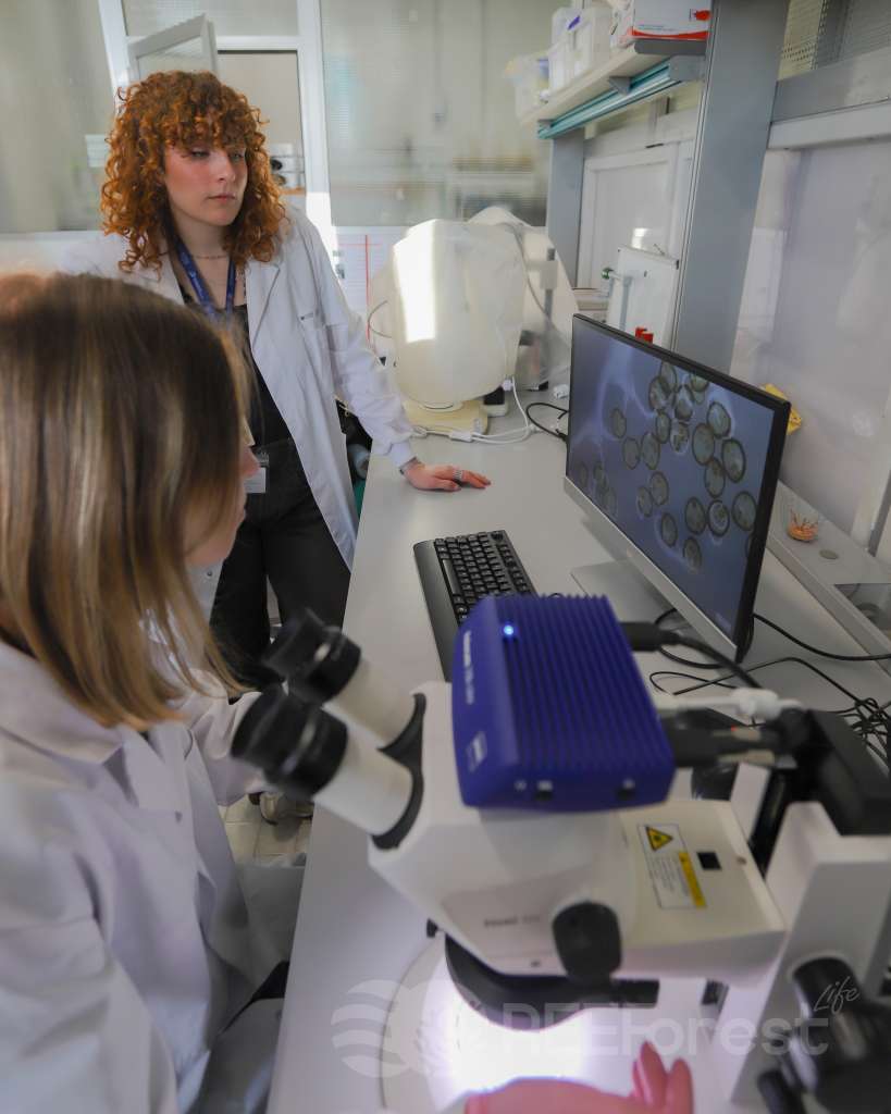
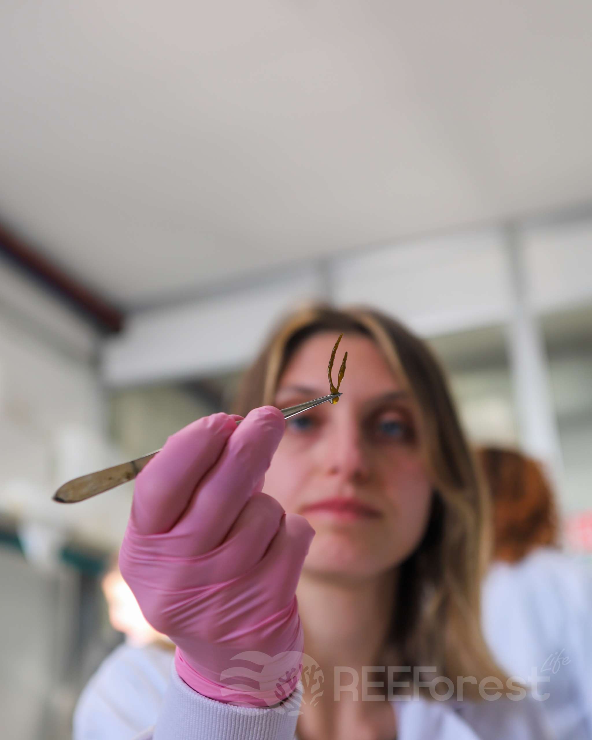
Starting at the end of winter, it is relatively easy to see without assistance the swellings at the end of the fronds which may indicate current fertility, but to be absolutely certain we must delve into the microscopic world.
Those swollen structures at the end of the fronds are the receptacles, the part of the algal thallus that contains the structures for reproduction.
Timing is essential: early or late harvesting can ruin the effort put into the laboratory cultures..
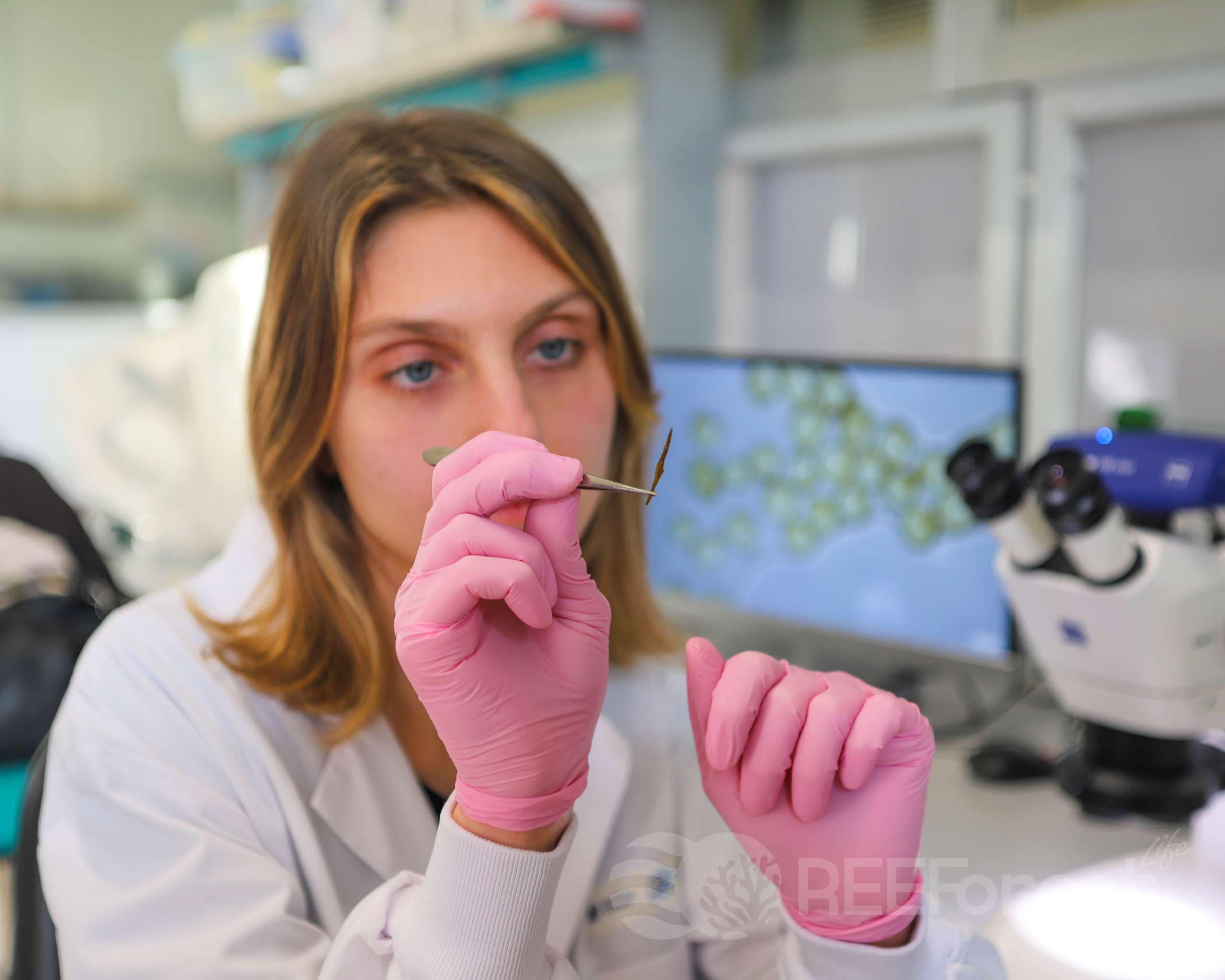
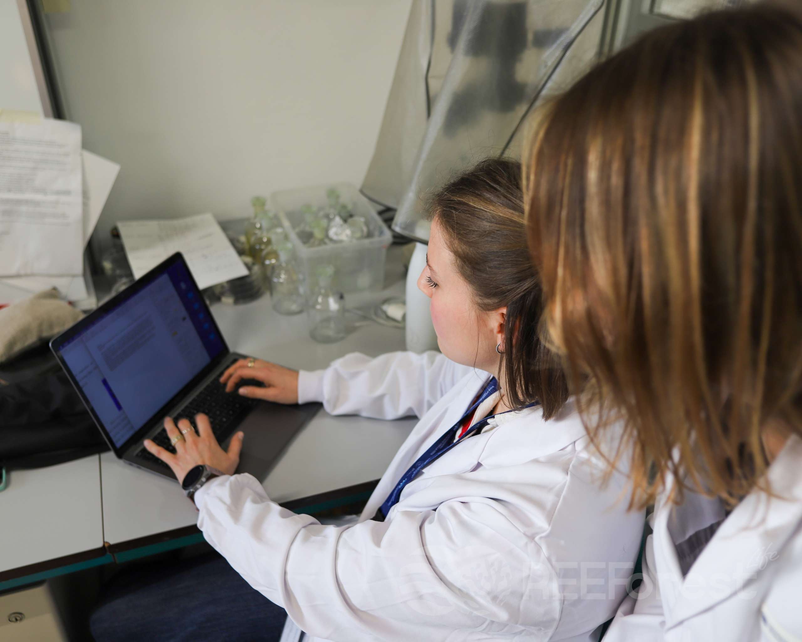
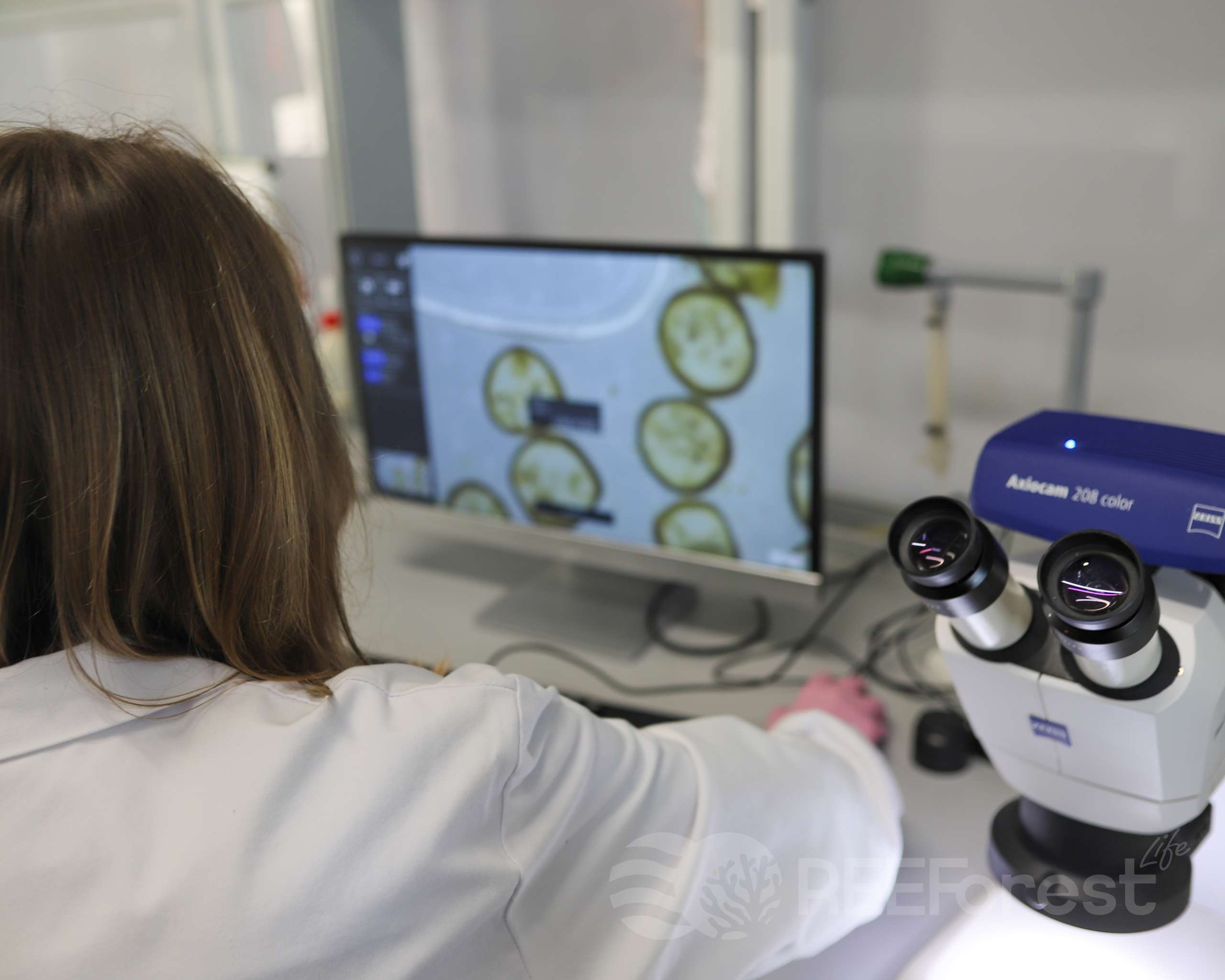
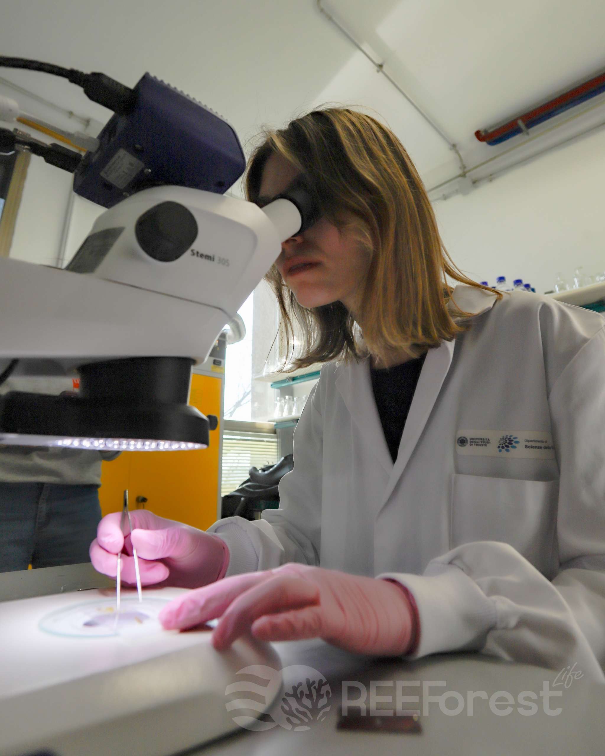
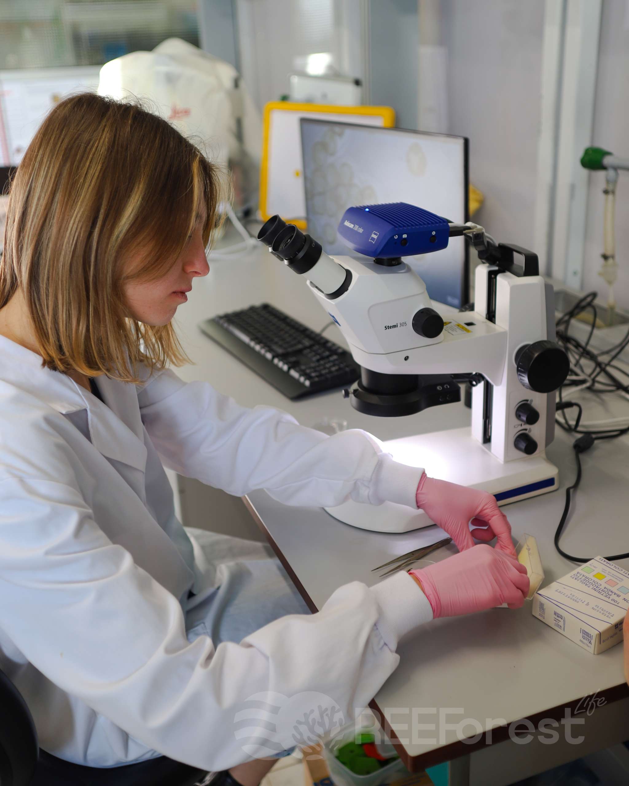
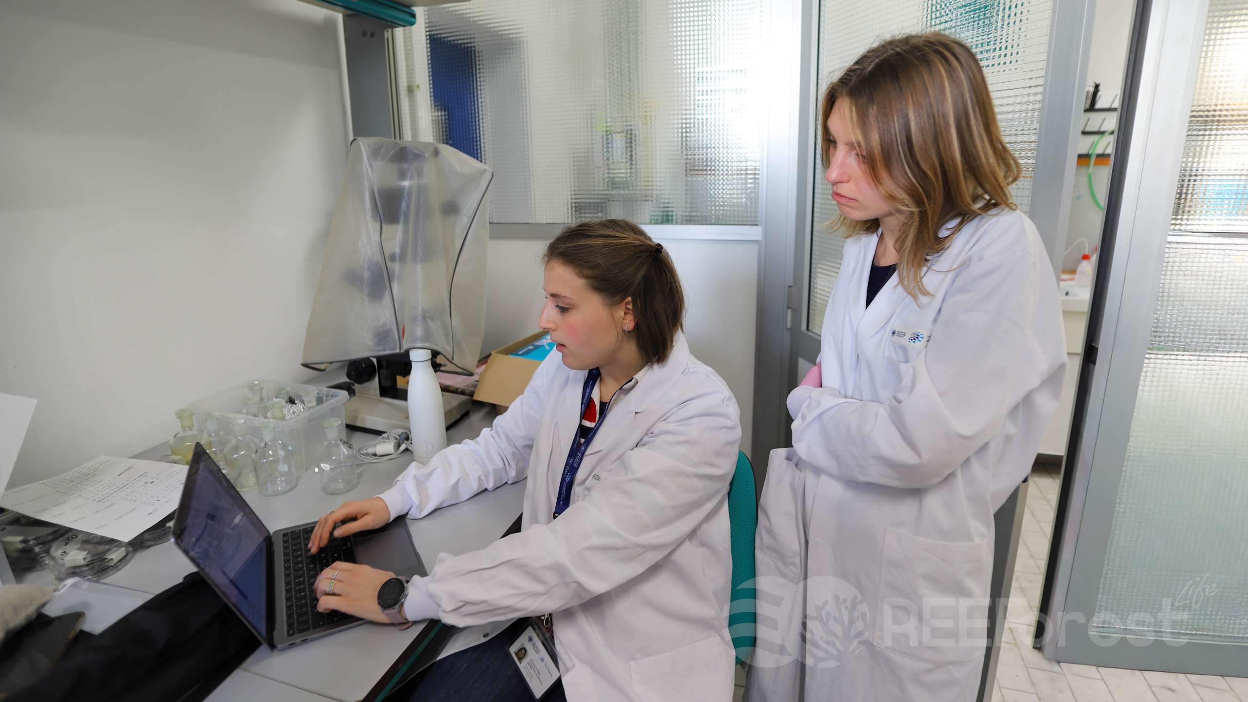
For this reason, before the fertile apices are harvested, our researchers assess the fertility of Cystoseira sensu lato species through visual analysis in the laboratory.
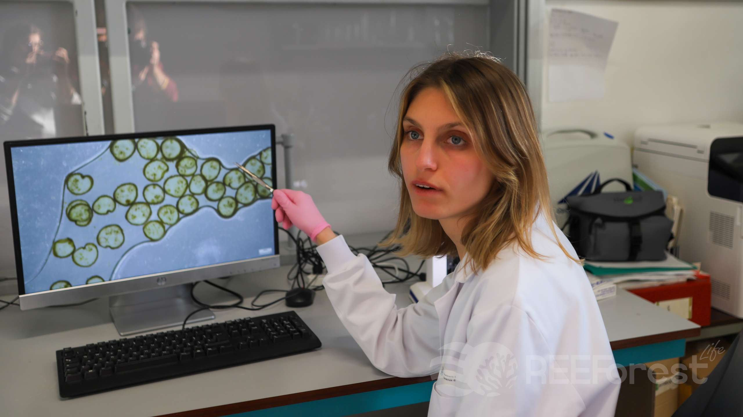
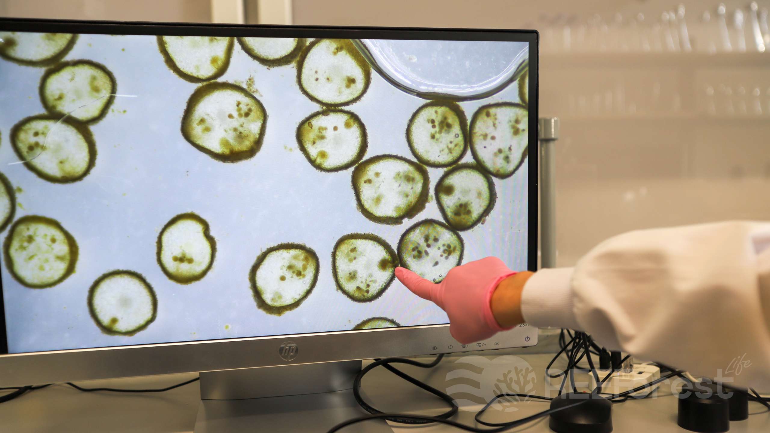
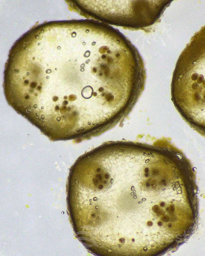
Pores called ostioles can be observed on the receptacles’ surface, each of which leads to a cavity called a conceptacle. These conceptacles host female (oogonia) and male (antheridia) gametangia on the inside. Cystoseira s.l. are hermaphroditic species: each thallus can host both female and male reproductive organs.
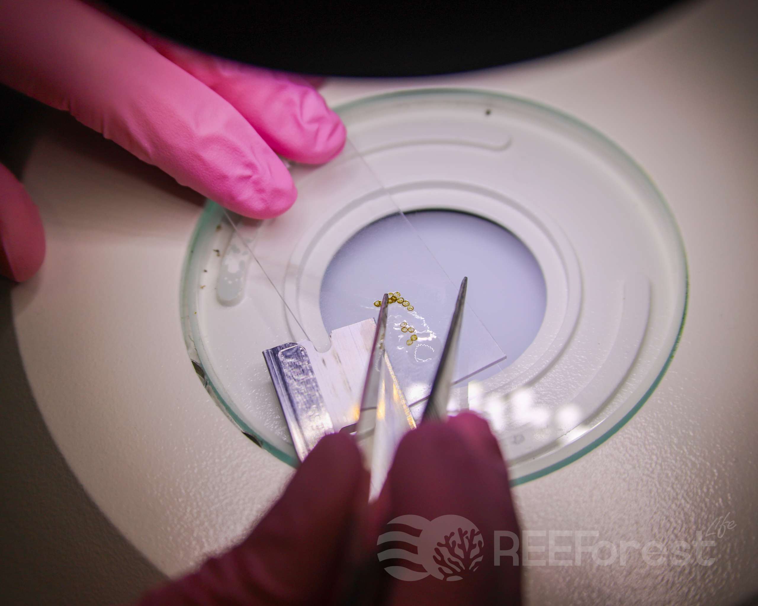
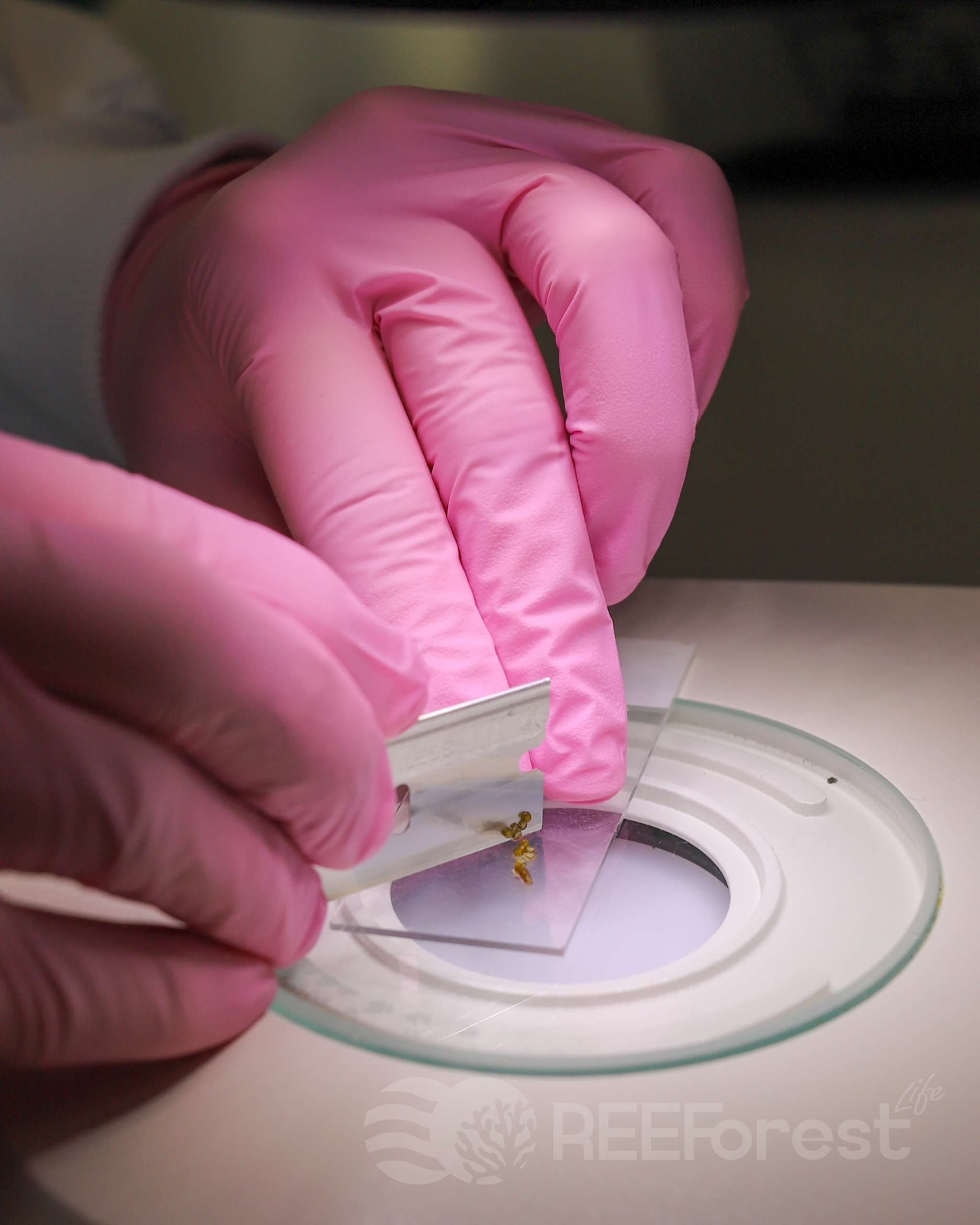
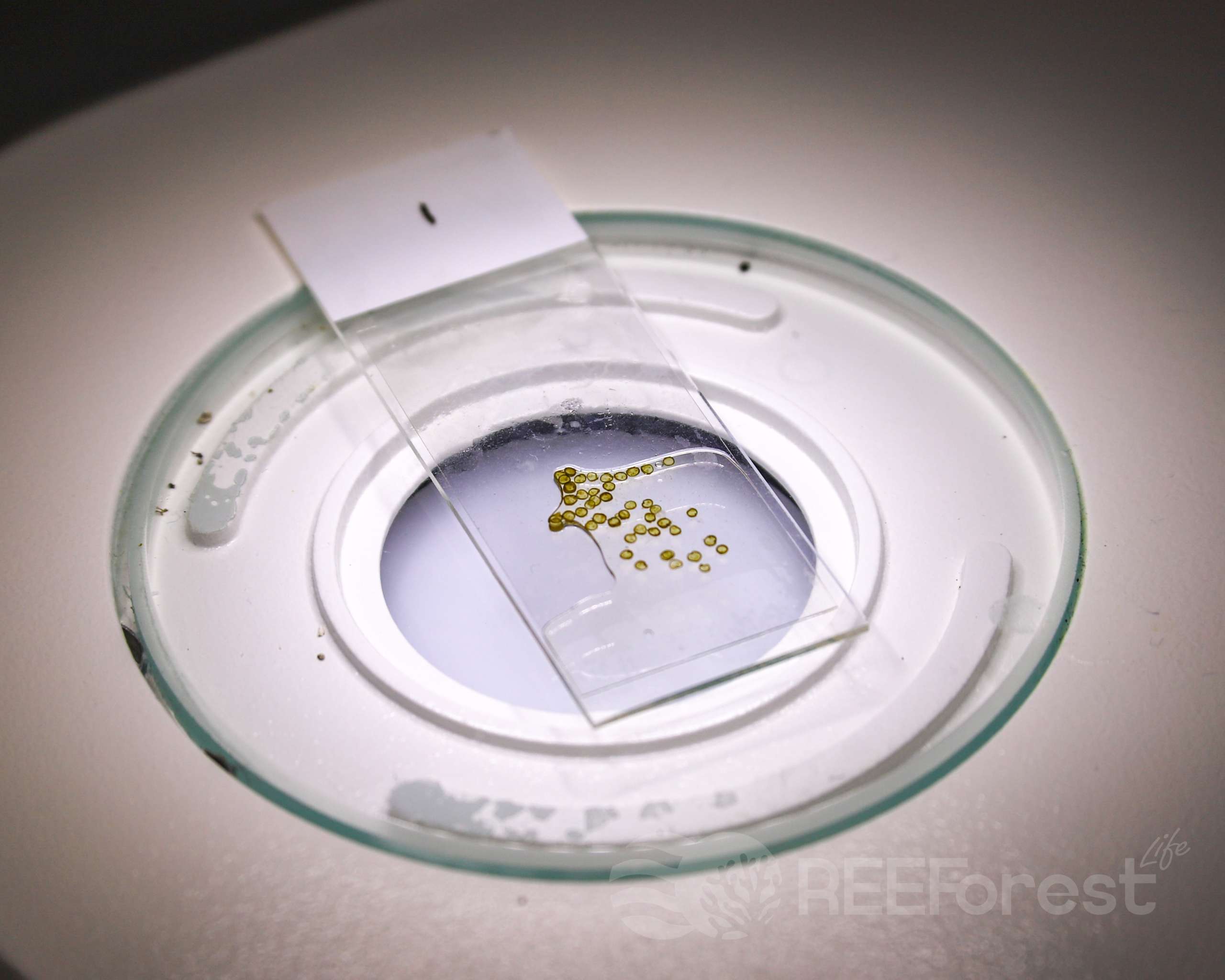
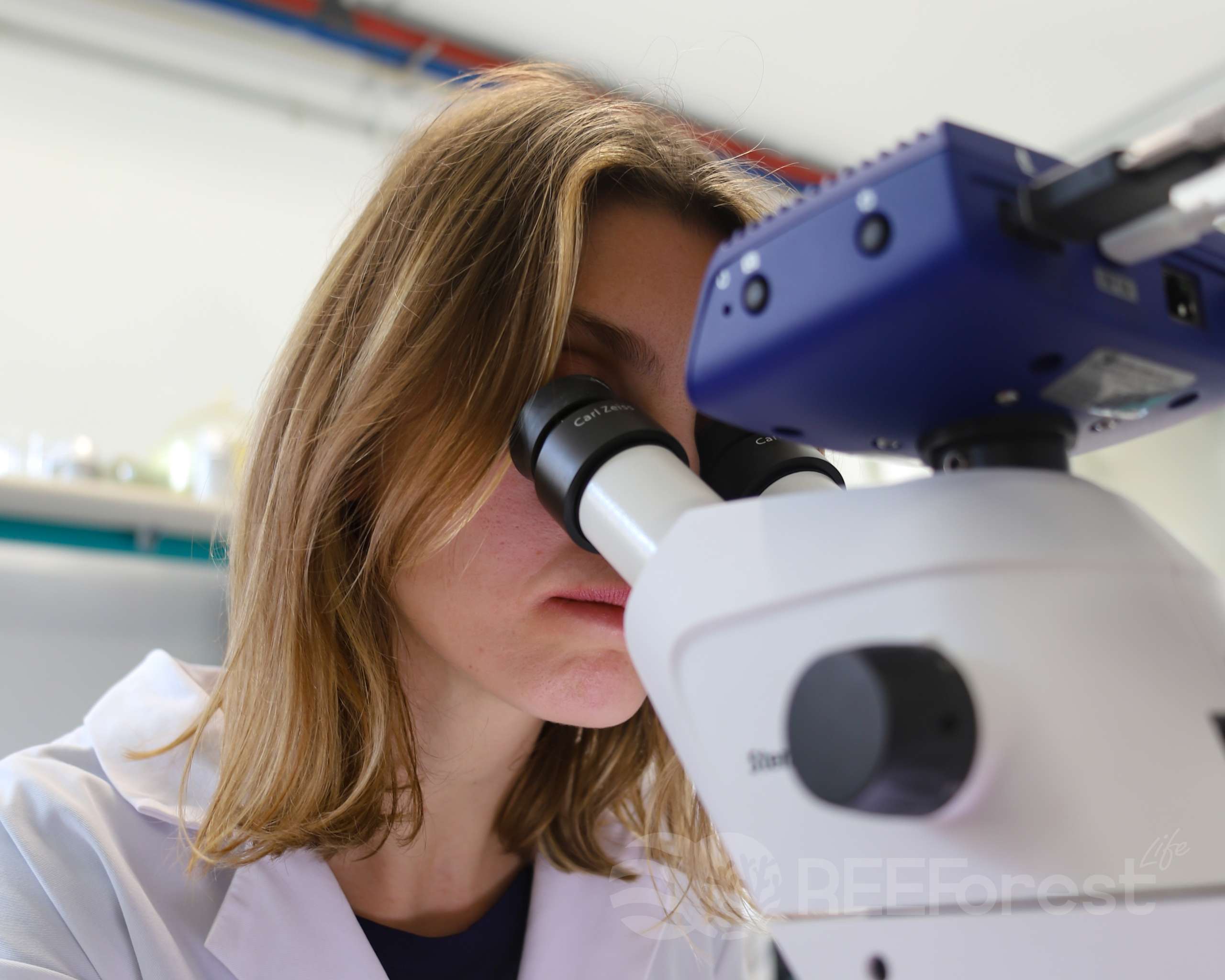
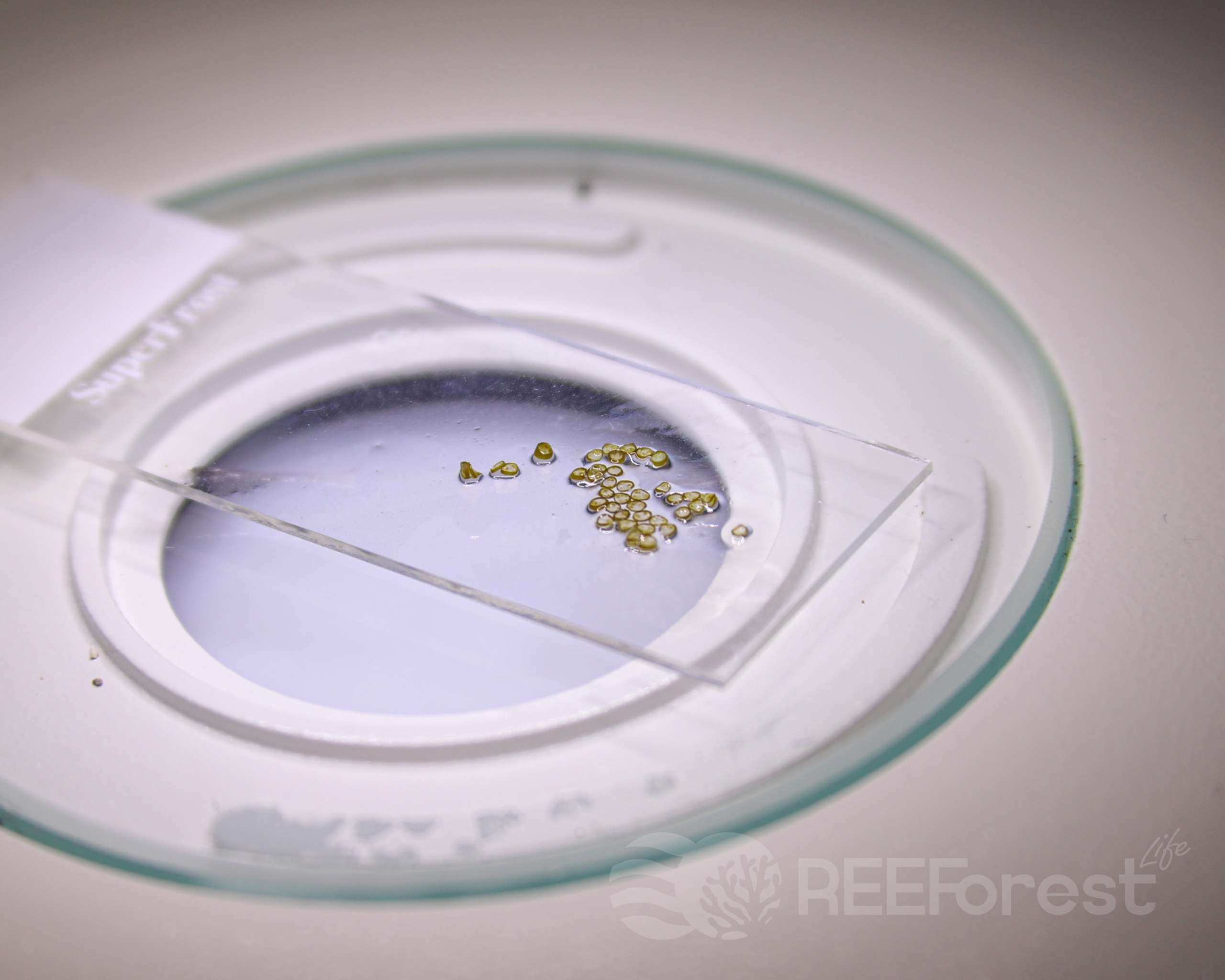
To assess the maturity stage of the sample, it is useful to cut thin sections of the receptacles and observe the conceptacles under a stereomicroscope, evaluating the number and size of the oogonia. The magnification required to appreciate the presence of gametes is 4x.
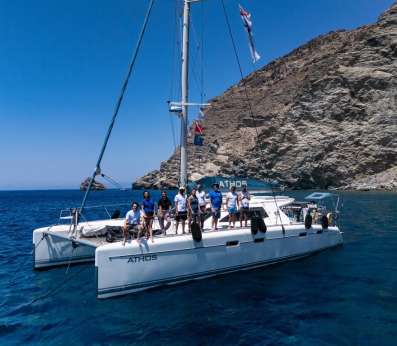
When the researchers will detect the optimal parameters indicating high fertility, the team will immediately leave for the collection site.
It should be noted that fertile apices are always collected in the area closest to the receiving site that still hosts a thriving and healthy population of Cystoseira s.l. The collection is carried out without damaging the thalli from which the fertile apices are detached. These fronds will release the embryos straight onto the clay discs where the new algae will grow.
The entire process follows the experience gained over the years to increase the chances of obtaining an abundant and resilient culture, to ensure that the stages following transplantation lead to efficient and long-lasting restoration.